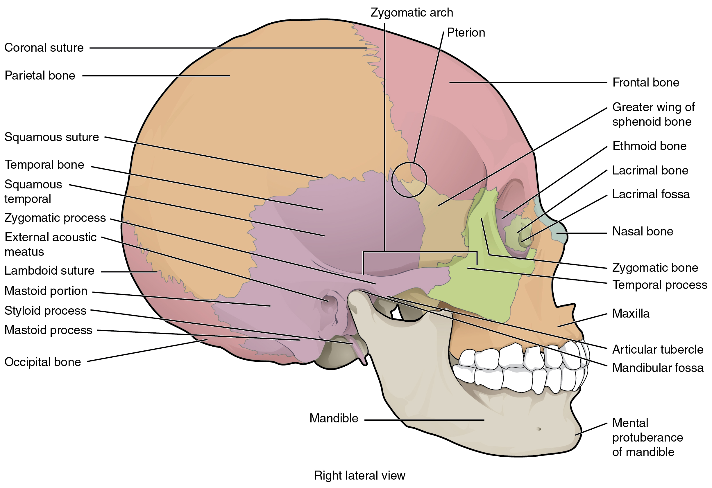
Back Of Head Skull Anatomy - Human Skull Anatomy Images, Stock Photos & Vectors ... - The neurocranium consists of the frontal, the ethmoid, the sphenoid, the occipital, and the paired temporal and parietal bones.
byAdmin•
0
Back Of Head Skull Anatomy - Human Skull Anatomy Images, Stock Photos & Vectors ... - The neurocranium consists of the frontal, the ethmoid, the sphenoid, the occipital, and the paired temporal and parietal bones.. They don't move and united into a single unit. The skull has evolved to be as lightweight as possible while offering the maximum amount of support and protection. A human skull is almost full sized at birth. It was then cleaned, adapted and polypainted this model is part of a comparison with the skull of a human. Let's get that view back.
If we just zoom in a bit, you can see something that's extending backwards from the palatine. The skull is a bone structure that forms the head in vertebrates. In the adult, the skull consists of 22 individual bones, 21 of which are immobile and united into a single unit. The skull begins to form prior to week 12 of embryogenesis. The skull anatomy has many functions aside from protecting the brain.
The Skull · Anatomy and Physiology from philschatz.com Bone that forms the forehead. In some back of the head skull deformities the contour deficiencies may extend up onto the top of the skull, most commonly into the parasagittal surfaces. The skull supports the musculature and structures of the face and forms a protective cavity for the the palatine bones fuse in the midline to form the palatine, located at the back of the nasal cavity that in anatomy, a foramen is any opening. Rectangular shaped bone on the sides of the head. For each skull base foramen, it is very important to remember its neurovascular relationships. Skull, skeletal framework of the head of vertebrates, composed of bones or cartilage, which form a unit that protects the brain and some sense organs. The cranium (skull) is the skeletal structure of the head that supports the face and protects the brain. The skull performs vital functions.
The cranium (skull) is the skeletal structure of the head that supports the face and protects the brain.
Let's get that view back. The skull is the skeleton of the head. The neurocranium consists of the frontal, the ethmoid, the sphenoid, the occipital, and the paired temporal and parietal bones. Anatomy of the skull and bones of cranium on medical illustrations. It offers protection to the brain, eye balls, inner ears, and nasal passages. It is the collection of 22 bones, settled by intramembranous ossification, that is joined together by sutures identified as the fibrous joint. Foramina inside the body of humans and other animals. It serves aesthetic purposes since it provides the visible appearance of the face. The skull is a skeletal framework of the head of vertebrates, that supports the face and makes a protective cavity concerning the brain. The 22nd bone is the mandible (lower jaw), which is the only moveable bone of the skull. The cranium and mandible was exported from ct data. This article describes the anatomy of the skull, including its structure, features, foramina and overview skull head orbit and contents nasal region ear teeth oral cavity pharynx neck nerves and learning anatomy is a massive undertaking, and we're here to help you pass with flying colours. Learn about anatomy skull with free interactive flashcards.
It supports and protects the face and the brain. Excluding ear ossicles, it is made of 22 bones. For each skull base foramen, it is very important to remember its neurovascular relationships. It is comprised of many bones, formed by intramembranous ossification, which are joined together by sutures (fibrous joints). The neurocranium consists of the frontal, the ethmoid, the sphenoid, the occipital, and the paired temporal and parietal bones.
MIDTERM - Anatomy & Physiology Bsc2085 with Ruiz at Miami ... from s3.amazonaws.com If we just zoom in a bit, you can see something that's extending backwards from the palatine. It is comprised of many bones, formed by intramembranous ossification, which are joined together by sutures (fibrous joints). In some back of the head skull deformities the contour deficiencies may extend up onto the top of the skull, most commonly into the parasagittal surfaces. However the eight bones that make up the cranium are not yet fused together. Let's get that view back. The cranium (skull) is the skeletal structure of the head that supports the face and protects the brain. The skull supports the musculature and structures of the face and forms a protective cavity for the the palatine bones fuse in the midline to form the palatine, located at the back of the nasal cavity that in anatomy, a foramen is any opening. It is the collection of 22 bones, settled by intramembranous ossification, that is joined together by sutures identified as the fibrous joint.
The skull is the bony skeleton of the head.
This anatomic region is complex and poses surgical challenges for otolaryngologists and neurosurgeons alike. It supports the structures of the face and provides a protective cavity for the brain. This means that the skull can flex and deform during birth, making it easier to deliver a baby through the narrow birth canal. Please feel free to download and print. However the eight bones that make up the cranium are not yet fused together. Let's get that view back. The cranium (skull) is the skeletal structure of the head that supports the face and protects the brain. These joints fuse together in adulthood. Excluding ear ossicles, it is made of 22 bones. In the adult, the skull consists of 22 individual bones, 21 of which are immobile and united into a single unit. The skull or known as the cranium in the medical world is a bone structure of the head. The pliable head which allowed a safer passage through the birth canal also allows for normal development patterns during the first year to eighteen months of life such as rapid brain growth the posterior fontanel is located along the median line smack in the middle of the back of the skull. The skull performs vital functions.
The skull includes the upper jaw and the cranium. Tables 2.1 and 2.2 triangles of the skull base. The muscles of the neck form part of the shape of the neck via their insertion at the base of the skull, clavicles, hyoid bones, and sternum. This means that the skull can flex and deform during birth, making it easier to deliver a baby through the narrow birth canal. Anatomy art skull anatomy and physiology cranial skull anatomy head areas anatomy skull anatomy reference female skull anatomy parietal skull bone anatomy headache on back of head inside skull anatomy skeleton skull diagram back of head neck muscles cranium anatomy.
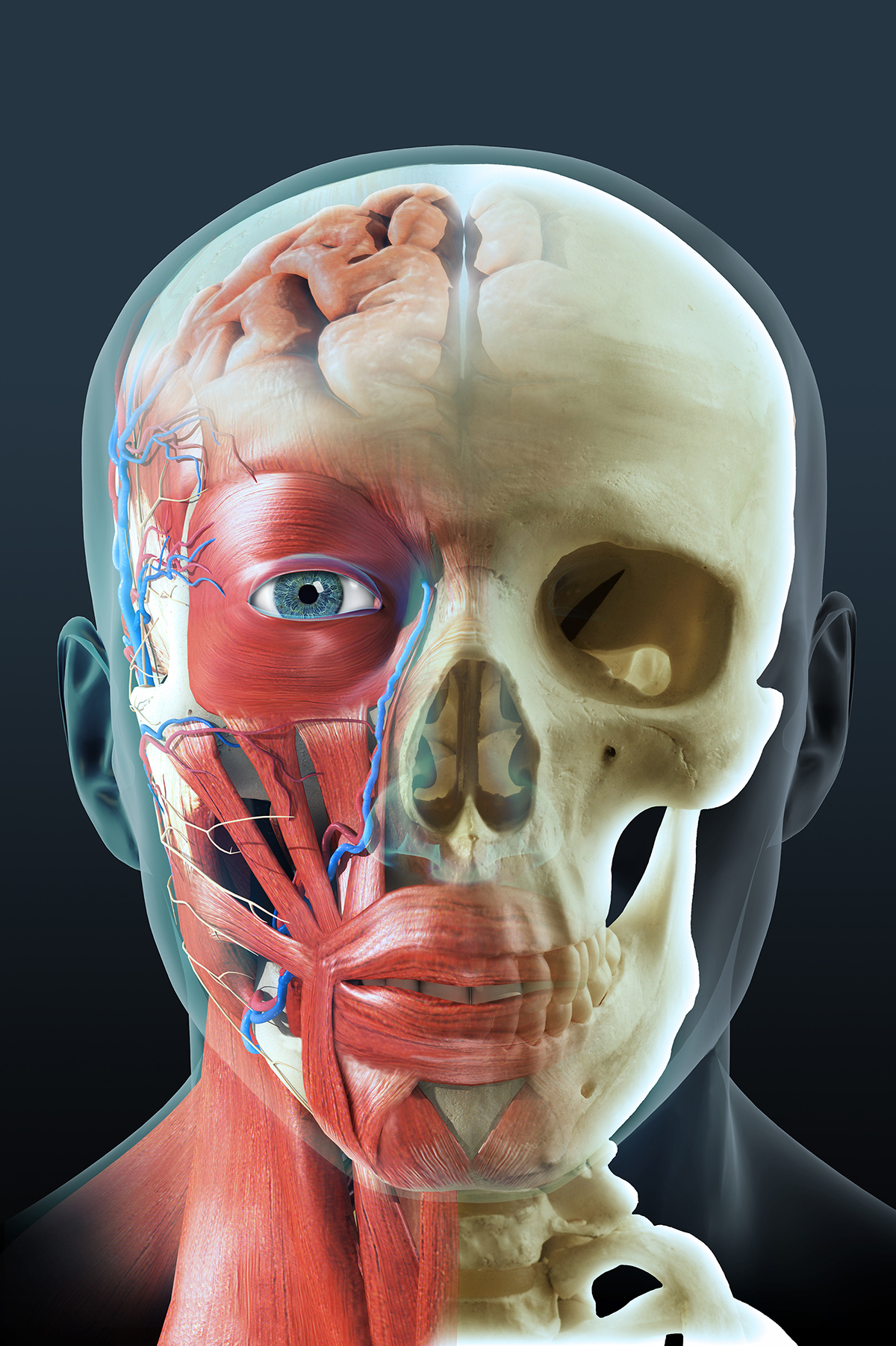
Anatomy of the head - AKYU from www.akyudesign.com The skull begins to form prior to week 12 of embryogenesis. Foramina inside the body of humans and other animals. It supports the structures of the face and provides a protective cavity for the brain. In some back of the head skull deformities the contour deficiencies may extend up onto the top of the skull, most commonly into the parasagittal surfaces. For each skull base foramen, it is very important to remember its neurovascular relationships. A human skull is almost full sized at birth. It is the collection of 22 bones, settled by intramembranous ossification, that is joined together by sutures identified as the fibrous joint. These joints fuse together in adulthood.
Anatomy of the head and neck.
These joints fuse together in adulthood. The skull begins to form prior to week 12 of embryogenesis. Cranial cavity , cranial sutures. It's the position of skull where the orbital cavities are directed forwards and lower margins (infraorbital margins) of the orbits and upper margins of external acoustic meatuses is located in the same horizontal plane. Anatomy of the head and neck. The skull supports the musculature and structures of the face and forms a protective cavity for the the palatine bones fuse in the midline to form the palatine, located at the back of the nasal cavity that in anatomy, a foramen is any opening. In order to be light, the skull is made up by flat and irregular bones, and has hollow spaces called the sinuses. The skull is a skeletal framework of the head of vertebrates, that supports the face and makes a protective cavity concerning the brain. It supports and protects the face and the brain. The sagittal suture is the line where the right and left parietal bone are in contact. It offers protection to the brain, eye balls, inner ears, and nasal passages. Bone that forms the forehead. It was then cleaned, adapted and polypainted this model is part of a comparison with the skull of a human.
It is the collection of 22 bones, settled by intramembranous ossification, that is joined together by sutures identified as the fibrous joint back of skull anatomy. The skull includes the upper jaw and the cranium.